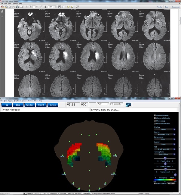
The following example shows an MRI of a client with a brain lesion that is detectable as a degeneration of the right insula. This was also detectable using sLORETA EEG imaging, by comparing the right and left insula in the beta band. As shown, the BrainAvatar sLORETA image also shows the reduced activity on the right side, confirming the MRI result. As an alternative application, this suggests that BrainAvatar sLORETA could be used as a screening, to indicate suggested areas of dysfunction, indicating the need for further study such as the more expensive MRI. This example shows that every voxel in BrainAvatar is converted to a quantity, the current-source density. This provides significant detail in space as well as time, when using this imaging approach. The current-source density data can also be used live for neurofeedback training, using any frequency bands or combination of frequency bands.
We would like to thank Thomas Feiner for providing this example.